Mechanosensitivity
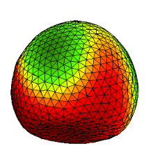
The responds of single cells to mechanical stimuli underly highly dynamic and sensitive mechanisms that maintain the proper functioning and development of various biological systems, such as mineralization of mussels[1], axon growth[2], and cardiac contraction and morphology[3, 4]. The mechanotransduction in cells can be processed by different complex chemo-mechanical and electro-mechanical coupling processes depending on the organism.
Here, we are interested in the dynamics and mechanisms of actin cortex sensing, force transmission and membrane & cytoskeletal proteins responses in electro-mechanical coupled cardiac tissue, which maintains the synchronization of tissue contraction and electrophysiological wave dynamics[3]. Other muscle cells are investigated to gain a fundamental understanding of the underlying processes and dynamics.
Quantitative and Computational Cell-Biology
Merging cell biology and systems biology was one of the initial drives to establish quantitative cell-biology using multivariate measurements, correlation analysis and causal inference that can be applied to learn the functional molecular causality underlying cellular activities across different scales[5]. The success of this approach have led over the years to an independent and interdisciplinary field, where mathematical and computational tools were used to enhance the understanding of various biological phenomena, such as the structural analysis of mechanosensitive cell responses[6], and multi-dimensional quantification and visualization of cell and tissue dynamics[7, 8].
Here, we aim to develop novel computational and analytical methods to extract and quantify morphological and functional dynamics in biological organisms on different length scales by interlinking biology, physics and computational science.
References
[1] V. Schönitzer, N. Eichner, H. Clausen-Schaumann and I. Weiss, Biochem. and Biophys. R. Comm. 415, 586, 2011
[2] D.E. Koser, et al., Nat. Neurosc. 19, 1592-1598, 2016
[3] M. Hörning, et al., Biophys. J. 102, 379-387, 2012
[4] M. Hörning and E. Entcheva, Book Chapter, Springer, 217, 237-258, 2015
[5] P. Liberali and L. Pelkmans, Nat. Cell Biol. 14, 1233, 2012
[6] M. Hörning, M. Nakahata, et al. Sci. Rep., 7, 7660, 2017
[7] M. Hörning, F. Blanchard, A. Isomura and Y. Yoshikawa, Sci. Rep. 7, 7757, 2017
[8] M. Hörning and T. Shibata, Biophys. J. 116, 2, 372-382, 2019


